Surgery to Repair Long-Gap Esophageal Atresia: Foker Process

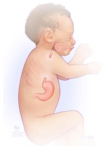
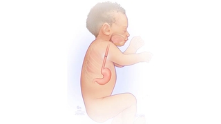
Esophageal atresia (EA) is a birth defect where there is a gap in the esophagus (the tube that connects the mouth to the stomach). In infants born with EA, the esophagus grows in two separate segments that do not connect. Surgery is needed to connect them so that a baby can swallow.
When the two segments of the esophagus are far apart, it is called a “long-gap” esophageal atresia. To repair a long-gap EA, a staged surgical process will be used. This procedure is called the “Foker process.” Surgeons carefully guide the growth of the two segments to bring them together.
What to expect during long-gap EA repair
Before surgery, a gastrostomy tube (G-tube) is placed surgically to feed your baby directly into the stomach. This allows for good growth before surgery.
The first stage of the Foker process involves placing stitches (also called sutures) near the ends of the two segments of the esophagus. These sutures are passed through the baby’s chest, and then tied in knots on the outside.
Approximately three times a week, surgeons carefully adjust these sutures. They first insert small tubes under the suture knots. Then, they gently apply tension to encourage the two ends of the esophagus to grow closer together. X-rays are used to monitor progress.
Throughout this process, your baby will be kept comfortable and safe. They will be sedated to help them feel more relaxed. They will be on a ventilator to help them breathe and will be kept very still to make sure the sutures stay secure.
Once the esophagus has grown enough so that the segments can reach each other, we can complete the repair. In this final stage, surgeons stitch the two ends together to create a functioning esophagus.

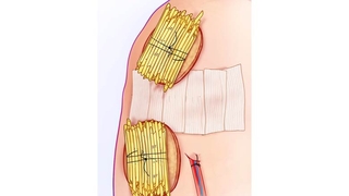
The upper and lower pouches of the esophagus are shown (inside the body) with sutures using a gentle pull or tension to encourage growth. The yellow tubes (outside the body) allow the surgeons to add tension as the esophagus grows to make the gap closer together.

The yellow tubes placed on top of the skin are used for adding gentle pull or tension to the pouches of the esophagus. The surgical incision is covered with white steri-strips to keep the area clean while it heals, and a small tube is placed to drain any fluid or air from the chest.

After the Foker process is complete, the two segments of the esophagus are surgically connected.
Recovery and follow-up care after surgery
Once your baby’s EA and/or tracheoesophageal fistula (TEF) are repaired, the most important next step is to allow time for healing.
Some special tests can be performed to be sure the repair is complete and successful. These tests include:
- Esophagram – This is a special study done using contrast or dye. The dye is either given by mouth or by a small feeding tube. It helps to show the shape of the esophagus and if there are any leaks and to make sure the esophagus has healed well.
- Esophagoscopy (EGD) – This test uses a small camera to view the esophagus. EGD studies will be repeated after esophageal repair to check on the healing of the esophagus. The EGD can also allow for gentle stretching (also called dilation) of the esophagus and collect tissue samples. Your baby’s GI doctor or endoscopist will help you plan for how often this test may be needed after surgery and after your baby is discharged home from the hospital.