Pediatric Pacemakers & Implantable Cardioverter Defibrillators (ICD)
What is an implanted pacemaker?
An implanted pacemaker is a small device used to treat patients with abnormally slow heart rhythms. The device is typically implanted under the skin. It produces an electrical signal that stimulates the heart and causes it to beat.
An implanted pacemaker may be used in situations where the heart's natural pacemaker (the sinoatrial node, or SA node) is not working properly, or when other parts of the cardiac electrical system are blocked.
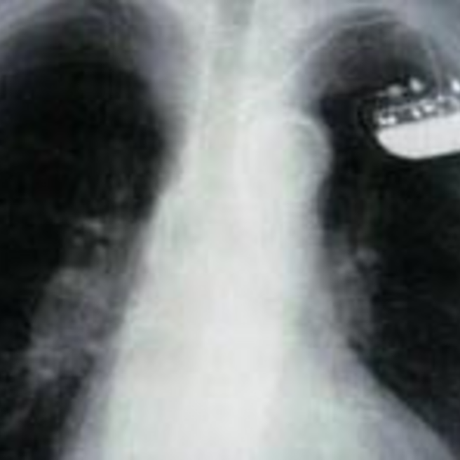
Children's pacemakers may be placed under the skin in one of several locations. Young children (infants, toddlers, preschool- and young school-aged children) often have the pacemaker generator placed in the abdomen. In older children and adolescents, the pacemaker generator is often placed in the chest, just under the collarbone.
What is an implantable cardioverter defibrillator (ICD)?
An implantable cardioverter defibrillator (ICD) is a small device used to treat patients who have, or are at risk of developing, abnormally fast heart rhythms that may lead to a cardiac arrest.
An ICD can detect these life-threatening heart rhythms almost immediately. When dangerously rapid heart rhythms persist and exceed a certain preprogrammed heart rate threshold, the ICD is able to deliver an electrical rescue shock to restore the normal heart rate. All ICDs are also pacemakers and are capable of pacing the heart when abnormally slow rhythms occur.
Why might a child require a pacemaker or an ICD?
Abnormal heart rhythms, or arrhythmias, impair the heart’s ability to effectively pump blood throughout the body. Slow rhythms, or bradyarrhythmias, reduce the amount of blood that is pumped out over time. Fast rhythms, or tachycardias, impair the heart’s ability to fill with blood. Both fast and slow heart rhythms can decrease cardiac output (the amount of blood that the heart pumps each minute), leading to symptoms like fatigue, exercise intolerance or fainting.
Pacemakers are generally recommended for patients with abnormally slow heart rhythms. When the heartbeat is too slow, the pacemaker will increase the heart rate and restore normal cardiac output. Slow heart rhythms may occur if the heart’s natural electrical system is damaged as a result of heart surgery, or because of rare inheritable cardiac diseases.
These slow heart rhythms may cause symptoms like fatigue, or they may limit the patient’s ability to exercise or participate in normal, everyday activities. Pacemakers may also be indicated for patients with abrupt pauses in the heart rhythm that lead to fainting.
When the heart is beating too fast, there is not enough time for the chambers to fill with blood between beats. A defibrillator can shock the heart when it’s beating too rapidly, which restores a normal rate and rhythm.
ICDs are usually recommended for patients at risk of abnormally rapid heart rhythms that may lead to cardiac arrest or sudden cardiac death. Some patients receive an ICD after they are rescued from a cardiac arrest.
Dangerously fast heart rhythms most often occur in patients who have scar tissue from prior heart surgery, or in patients with diseases of the heart muscle known as cardiomyopathies. Certain cardiac electrical diseases like long QT syndrome, Brugada syndrome, or inheritable forms of ventricular tachycardia may also be associated with life-threatening rapid heart rhythms.
Occasionally, patients with significant heart failure may benefit from ICD placement to protect them from sudden death while they are awaiting heart transplantation.
What are the components of a permanent pacemaker/ICD?
A permanent pacemaker has two components, including:
- A pulse generator which has a sealed lithium battery and an electronic circuitry package. The pulse generator produces the electrical signals that make the heart beat. Many pulse generators can also interpret signals that are sent by the heart itself.
- One or more wires (also called leads). Leads are insulated, flexible wires that conduct electrical signals to the heart from the pulse generator. The leads may also relay signals from the heart to the pulse generator. One end of the lead is attached to the pulse generator and the other end of the lead is positioned in the heart. Pacemakers may have leads that connect to the atrium (the upper chamber of the heart) the ventricle (the lower chamber of the heart) or both. In the case of a biventricular pacemaker, leads are placed in both ventricles.
Pacemakers can "sense" when the heart's natural rate falls below the rate that has been programmed into the pacemaker's circuitry.
ICD systems are made up of a generator and one or more leads, just like a pacemaker. ICD leads are typically larger than pacemaker leads and include an additional component that enables the device to deliver a high-voltage shock.
How are pacemakers and ICDs implanted?
Pacemakers and ICDs are inserted in the hospital, and children usually receive general anesthesia during the procedure. Patients are usually kept overnight for monitoring after a new device is inserted.
There are two basic kinds of pacemaker systems: epicardial and transvenous.
- Epicardial pacemakers and defibrillators have leads that are attached to the outside of the heart. They are generally placed in smaller children (infants to younger school-age). The device itself is usually placed in the belly, as there is more room there and the device is larger in proportion to the size of the child. The leads must be sewn onto the surface of the heart, which involves opening the chest. The procedure is performed by a pediatric cardiac surgeon in an operating room, and children are always kept in the hospital overnight (and sometimes for several days). Usually a temporary chest tube is placed to allow fluid to drain after the procedure.
- Transvenous pacemakers and defibrillators have leads that course through the veins and are fixed to the inside of the heart. They are generally an option for larger children and adolescents. These devices can be implanted without opening the chest, and the procedure is typically less invasive than placement of an epicardial device. The procedure is performed by a cardiologist and/or cardiac surgeon in the catheterization lab. A needle is inserted into the vein under the clavicle and the pacemaker lead is placed in the heart through a large IV or intravenous sheath. Fluoroscopy (continuous x-ray) is used during the procedure to visualize the position of the lead inside the heart. The lead is then attached to the device, which is placed underneath the skin and outside of the ribs. Children are kept in the hospital overnight after a new transvenous implant, but the vast majority are able to go home the following day.

After a device is implanted, patients receive an identification card from the manufacturer that includes information about the specific model of pacemaker and the serial number. The device can be reprogrammed and checked regularly by a cardiologist in the office. Limited device checks can also be done from home over the phone.