
Those with Down syndrome have increased joint mobility and laxity. They are able to move at their joints in extreme ways. This is one reason they are so flexible and are often referred to as “double jointed.” They don’t actually have double joints, but have excessive movement and ligamentous laxity.
Babies are especially flexible because their muscles, joints and bones have not developed to provide additional stability. Joints and bones develop as a person grows. Some of the contributing factors to this excessive movement include low muscle tone (hypotonia), decreased muscle strength and stretchy ligaments (or ligamentous laxity). Ligaments are bands of connective tissue that connect two bones or cartilage. This article explains ligamentous laxity of the foot, ankle and spine.
Understanding Joint Mobility and Stability
Joint mobility is the amount of movement that exists at two articulating surfaces before movement is restricted by surrounding tissues — ligaments, tendons, muscles, etc. Joint stability is the ability to control joint movement. We need both mobility and stability at each joint for proper movement and activity.

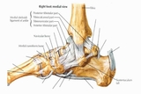
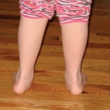
There are 28 bones and 30 joints and more than 100 tendons, muscles and ligaments in the foot and ankle, making movement at the foot and ankle complex. (See Figure 1.) Laxity of the ligaments at the ankle and foot is one reason those with Down syndrome stand with flat feet. We refer to this foot position as pronation. A pronated foot (see Figure 2) does not have the support to maintain adequate balance and stability. Overpronation also results in increased stress at the knee joint, which can lead to medial knee and low back pain as a person ages.

You may notice a person turns their feet out to the side or grips with their toes for balance when they have loose ligaments. Callouses, metatarsus adductus, decreased ankle mobility and pain are other compensations that may occur. Therapists frequently recommend orthotics or braces to support the foot in the absence of adequate ligament support.
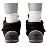
The goal of wearing an orthotic is to provide support and optimal alignment to the muscles and joints (see Figure 3) while allowing movement. No matter the exact type of orthotic your child wears (see Figure 4), it should provide the ability to move with the correct motor pattern, a more efficient gait pattern and improved balance.
Understanding Risks of Spine Instability

Ligaments of the spine may also be loose, which can lead to scoliosis and neck instability. This neck instability is commonly referred to as atlanto-axial instability.
The first vertebrae or bone in the neck is the atlas (C1) and the second is the axis (C2). The ligamentous laxity places those with Down syndrome at risk for neck injury. Because of their increased ligamentous laxity of the neck, the Academy of Pediatrics recommends that those with Down syndrome not participate in activities — such as football, head butting in soccer, diving or using a trampoline — that may result in neck trauma. Neck vertebrae may move or slide, which can lead to damage to the spinal column.
Scoliosis may occur if the bones or vertebrae in the back are not straight and a curve is present. This occurs in approximately 7% to 9% of those with Down syndrome. Screening for scoliosis is important. Your child will receive a referral to Orthopaedics if we detect a curve. The orthopedic specialist may order X-rays and determine if the curve requires bracing, monitoring or surgery.
Individuals’ Laxity Differs
Every person with Down syndrome displays a different degree of joint ligamentous laxity. Sometimes, ligaments and muscles become tighter as a person grows. Bone grows and ligaments and muscles stretch to accommodate new bone length.
We cannot directly change the amount of ligamentous laxity with exercise and therapy, but we can improve motor control, balance and strength, which may also improve stability around a joint. Bracing, kinesio taping and orthotics can also provide external support for individuals with ligamentous laxity.
Physical therapists are movement specialists who can assist those with Down syndrome with movement, mobility, strength and alignment. The goal of physical therapy is to promote a healthy, active lifestyle while preventing orthopedic impairments, pain and immobility, which can be a result of ligamentous laxity.
References
Gupton M, Terreberry RR. Anatomy, bony pelvis and lower limb, calcaneus. StatPearls website. https://www.ncbi.nlm.nih.gov/pubmed/30137829. Updated December 19, 2018. Accessed February 5, 2020.
Bull M, Committee on Genetics. Health supervision for children with Down syndrome. Pediatrics. 2011;128(2):393-406.
Featured in this article
Specialties & Programs

Those with Down syndrome have increased joint mobility and laxity. They are able to move at their joints in extreme ways. This is one reason they are so flexible and are often referred to as “double jointed.” They don’t actually have double joints, but have excessive movement and ligamentous laxity.
Babies are especially flexible because their muscles, joints and bones have not developed to provide additional stability. Joints and bones develop as a person grows. Some of the contributing factors to this excessive movement include low muscle tone (hypotonia), decreased muscle strength and stretchy ligaments (or ligamentous laxity). Ligaments are bands of connective tissue that connect two bones or cartilage. This article explains ligamentous laxity of the foot, ankle and spine.
Understanding Joint Mobility and Stability
Joint mobility is the amount of movement that exists at two articulating surfaces before movement is restricted by surrounding tissues — ligaments, tendons, muscles, etc. Joint stability is the ability to control joint movement. We need both mobility and stability at each joint for proper movement and activity.

There are 28 bones and 30 joints and more than 100 tendons, muscles and ligaments in the foot and ankle, making movement at the foot and ankle complex. (See Figure 1.) Laxity of the ligaments at the ankle and foot is one reason those with Down syndrome stand with flat feet. We refer to this foot position as pronation. A pronated foot (see Figure 2) does not have the support to maintain adequate balance and stability. Overpronation also results in increased stress at the knee joint, which can lead to medial knee and low back pain as a person ages.

You may notice a person turns their feet out to the side or grips with their toes for balance when they have loose ligaments. Callouses, metatarsus adductus, decreased ankle mobility and pain are other compensations that may occur. Therapists frequently recommend orthotics or braces to support the foot in the absence of adequate ligament support.
The goal of wearing an orthotic is to provide support and optimal alignment to the muscles and joints (see Figure 3) while allowing movement. No matter the exact type of orthotic your child wears (see Figure 4), it should provide the ability to move with the correct motor pattern, a more efficient gait pattern and improved balance.
Understanding Risks of Spine Instability

Ligaments of the spine may also be loose, which can lead to scoliosis and neck instability. This neck instability is commonly referred to as atlanto-axial instability.
The first vertebrae or bone in the neck is the atlas (C1) and the second is the axis (C2). The ligamentous laxity places those with Down syndrome at risk for neck injury. Because of their increased ligamentous laxity of the neck, the Academy of Pediatrics recommends that those with Down syndrome not participate in activities — such as football, head butting in soccer, diving or using a trampoline — that may result in neck trauma. Neck vertebrae may move or slide, which can lead to damage to the spinal column.
Scoliosis may occur if the bones or vertebrae in the back are not straight and a curve is present. This occurs in approximately 7% to 9% of those with Down syndrome. Screening for scoliosis is important. Your child will receive a referral to Orthopaedics if we detect a curve. The orthopedic specialist may order X-rays and determine if the curve requires bracing, monitoring or surgery.
Individuals’ Laxity Differs
Every person with Down syndrome displays a different degree of joint ligamentous laxity. Sometimes, ligaments and muscles become tighter as a person grows. Bone grows and ligaments and muscles stretch to accommodate new bone length.
We cannot directly change the amount of ligamentous laxity with exercise and therapy, but we can improve motor control, balance and strength, which may also improve stability around a joint. Bracing, kinesio taping and orthotics can also provide external support for individuals with ligamentous laxity.
Physical therapists are movement specialists who can assist those with Down syndrome with movement, mobility, strength and alignment. The goal of physical therapy is to promote a healthy, active lifestyle while preventing orthopedic impairments, pain and immobility, which can be a result of ligamentous laxity.
References
Gupton M, Terreberry RR. Anatomy, bony pelvis and lower limb, calcaneus. StatPearls website. https://www.ncbi.nlm.nih.gov/pubmed/30137829. Updated December 19, 2018. Accessed February 5, 2020.
Bull M, Committee on Genetics. Health supervision for children with Down syndrome. Pediatrics. 2011;128(2):393-406.
Contact us
Trisomy 21 Program